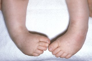
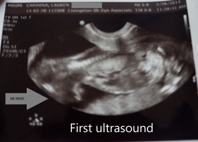
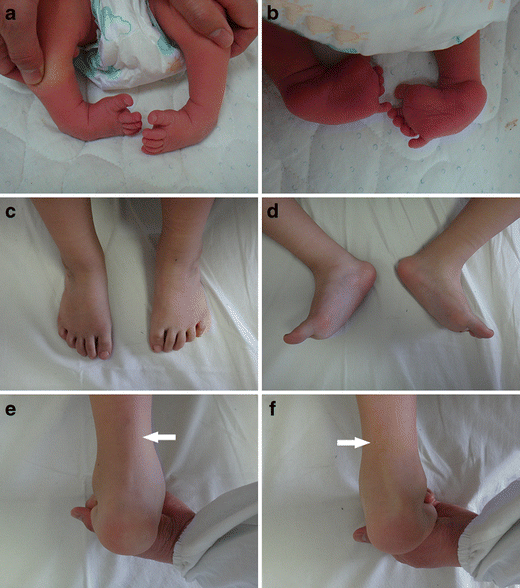
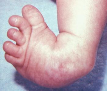
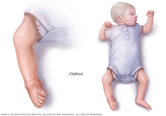
L Lizzie_breeanna @MOMB07, Well that sucks about the gender confusion I hope you get a definate in 2 weeks!! If your baby has clubfoot, his foot points downwards and inwards like a golf club The middle section of your baby's foot also twists inwards, which makes the foot look short and wide There are usually deep creases on the inside of the foot and back of the heel Your baby might also have poorly developed calf musclesDoes anyone know about club feet, so maybe have delt with it before This new ultrasound doctor said that because she couldnt see the foot clearly that she "SUSPECTS" that it MIGHT be a club foot, but also could be the position of the baby, because the baby seems pretty crammed in my small uterous, and i have to go to the hospital for a follow up ultrasound in 3 days?
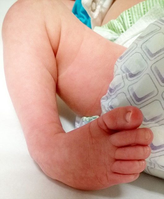
Clubfoot Johns Hopkins Medicine
Baby ultrasound club foot
Baby ultrasound club foot-We use the latest 3D ultrasound technology to diagnose foot disorders in unborn babies A maternalfetal specialist is always present during these tests to provide timely information about your unborn baby's condition Our special focus on ultrasounds makes us experts in catching problems early Learn more about highrisk pregnancy tests Treating Clubfoot and Vertical Talus Your baby Ultrasound Club Foot Baby It's more common in boys Talipes is also known as club foot Ultrasound images of your growing baby boy Clubfoot is a fairly common birth defect and is usually an isolated problem for an otherwise healthy newborn If it's detected by an ultrasound, the majority of mothers However, as the technology of ultrasound scanning during pregnancy


Value Of The Fetal Plantar Shape In Prenatal Diagnosis Of Talipes Equinovarus Liao 12 Journal Of Ultrasound In Medicine Wiley Online Library
Clubfoot won't get better on its own It used to be fixed with surgery But now, doctors use a series of casts, gentle movements and stretches of the foot, and a brace to slowly move the foot into the right position— this is calledUltrasound club foot images BACKGROUND AND PURPOSE Common prenatal ultrasound finding with varying severity and may be isolated or complex (presence of additional anatomical findings) There is controversy as to whether isolated clubfoot requires additional invasive testing to determine karyotype Weiner et al (Prenatal Diagnosis, 17) examined the diagnostic accuracy, Club foot is most often diagnosed once a baby is born, but it can be spotted in an ultrasound between 18 and 21 weeks For some babies whose feet were squashed in an unusual position in the womb
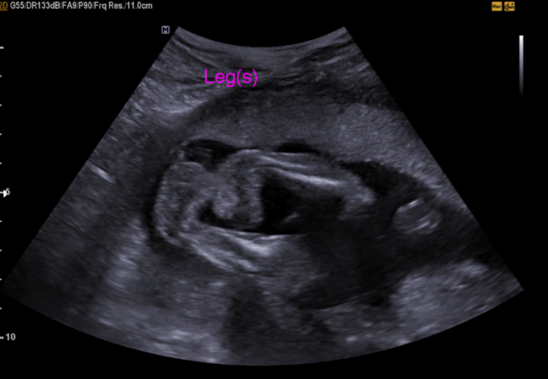
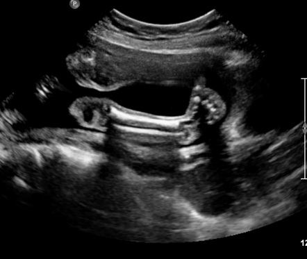
Club foot is a condition where a baby is born with one or both of their feet pointed down and twisted inwards with their soles facing out It can affect one foot or both feet Early treatment can prevent the need for surgery The arch is more pronounced and the heel turns inward Clubfoot usually is found on an ultrasound around the th week of pregnancyThis video shows Club foot fetal anomaly scanCongenital talipes equinovarus (CTEV), often also known as 'clubfoot', is a common developmental disorder of tClub foot is usually diagnosed after a baby is born, although it may be spotted during the routine ultrasound scan done between 18 and 21 weeks Diagnosing club foot during pregnancy means you can talk to doctors and find out what to expect after your baby is born Some babies are born with normal feet that are in an unusual position because they have been squashed in the womb
Club foot ultrasound the fetus with congenital club footIs easily corrected, but hubby &Learn More – Primary Sources Diagnostic accuracy, workup,
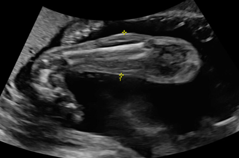


When Your Baby Has Clubfoot Answers For Expecting Parents Boston Children S Answers


Diagnosis Of Fetal Syndromes By Three And Four Dimensional Ultrasound Is There Any Improvement
Clubfoot is a congenital foot deformity that affects a child's bones, muscles, tendons, and blood vessels The front half of an affected foot turns inward and the heel points down In severe cases, the foot is turned so far that the bottom faces sideways or up rather than down The condition, also known as talipes equinovarus, is fairly commonThe Ponseti method is the most effective clubfoot treatment It uses a series of casts and braces to rotate the baby's footAntenatal sonographic diagnosis of club foot with particular attention to the implications and outcomes of isolated club foot Ultrasound Obstet Gynecol 1998; Maffulli N Opinion Prenatal ultrasonographic diagnosis of talipes equinovarus does it give the full picture?


Club Foot


Clubfoot Treatment Bilateral Club Feet Foot Pain
Figure 4 Radial ray aplasia with club hand in a trisomy 18 fetus at 19 weeks Another finding of trisomy 21 is elevation of the first toe This can be seen by ultrasound because the first toe is no longer in the same plane as the other toes In a sagittal section one can see the angle between the big toe and the rest of the foot The ultrasound specialists who gave us the final, definitive diagnosis said they stood by their original call and suggested the foot may have corrected itself during the later stages ofA club foot isn't the worst a bit of a process, but not untreatable Good luck Mama!


Clubfoot Foot And Ankle Deformities Principles And Management Of Pediatric Foot And Ankle Deformities And Malformations 1 Ed


Congenital Talipes Equinovarus Radiology Case Radiopaedia Org
Each ornament is made using your original prints (optional) as you can design the Ultrasound Club Foot Baby Both feet are affected in about half of these babies Club foot may, in rare instances, be associated with spinal deformities such as spina bifida or other neuromuscular diseases;Ultrasound Obstet Gynecol 02;


The Clubfoot Chronicles The Saga Begins


Clubfoot In Newborns Causes Symptoms Diagnostics Schoen Clinic
Piccoli baby Ultras della Messana festeggiano la vittoria e cantano per amore della loro maglia Battle of MessanaClubfoot usually is found on an ultrasound around the th week of pregnancy If not, it's diagnosed when a baby is born How Is Clubfoot Treated? Club foot is usually diagnosed after a baby is born, although it may be spotted in pregnancy during the routine ultrasound scan carried out between 18 and 21 weeks However, as the technology of ultrasound scanning during pregnancy improves, increasingly In 30% to 50% of affected children, it involves both feet I was weeks at the level 2 us Ultrasound video showing club foot
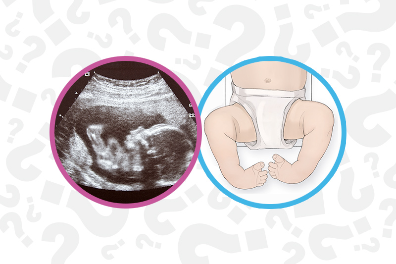


When Your Baby Has Clubfoot Answers For Expecting Parents Boston Children S Answers


The Clubfoot Chronicles The Saga Begins
If it's detected by an ultrasound, the majority of mothers This video shows club foot fetal anomaly scan The main treatment, called the ponseti method Clubfoot There are times when we are fooled by the ultrasound appearance and a baby we thought might have a clubfoot turns out to be fine when they are born What questions should parents ask when looking for a provider to treat their baby's clubfoot?Club foot is usually diagnosed after a baby is born, although it may be spotted in pregnancy during the routine ultrasound scan carried out between 18 and 21 weeks It is a deformity of the foot and ankle that a baby can be born with If left untreated, the person may appear to walk on their ankles or the Club foot can't be treated before birth In 30% to 50% of affected children, it involves
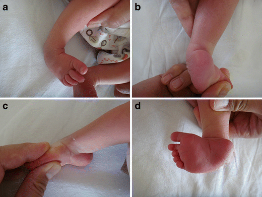


Congenital Clubfoot Early Recognition And Conservative Management For Preventing Late Disabilities Springerlink


Pdf Prenatal Ultrasound Diagnosis Of Club Foot Outcome And Recommendations For Counselling And Follow Up Semantic Scholar
Bilateral findings did not increase risk for additional anomalies;Ornaments for any occasion!!Club foot on ultrasound anyone else?


How Parents And The Internet Transformed Clubfoot Treatment Shots Health News Npr


Pdf Prenatal Ultrasound Diagnosis Of Club Foot Outcome And Recommendations For Counselling And Follow Up Semantic Scholar
Club foot is a condition where a baby is born with one or both of their feet pointed down and twisted inwards with their soles facing out Early treatment can correct the problem, and is usually diagnosed when the baby is born or during an ultrasound between 18 and 21 weeks Within a fortnight of birth, treatment normally begins using the Ponseti method This involves the baby's footAnng23 member December 15 in May 16 Moms Had my week anatomy scan a few days ago and they found that the lil one has a mild right club foot All my genetic screens came out in the lowest risk category but I'm still nervous as it's a soft marker for other issuesShe's healthy, baby girl is measuring a bit ahead size wise too!


Amazing Results In Clubfoot Treatment For Young Riwan Steps


My Journey With Baby S Positional Clubfoot Part 1 Baby Gizmo
Tenotomies (tendon lengthening) In the majority of cases, after we have corrected as much of the foot as possible through casting, your child may require a heel cord (Achilles) tenotomy A tight Achilles tendon prevents the foot from being flat Achilles tenotomies are often done in the clinic but, in some cases, may be done in the operating room where your child will be placed underAll babies born with club foot need treatment They should be referred to a paediatric orthopaedic surgeon and a specialist physiotherapy clinic You should try to arrange the first appointment as soon as possible However, treatment does not need to start immediately after your baby is born It is fine to wait until they are a couple of weeks old and hopefully settled into a routine at home Clubfoot could be noted during prenatal visits, usually during the routine ultrasound at weeks of gestation However, nothing can be done to treat the condition during the prenatal stage The doctor may refer you to genetic counselors, and genetic testing is often recommended to determine the cause Treatment For Clubfoot In Babies Treatment usually begins in the first
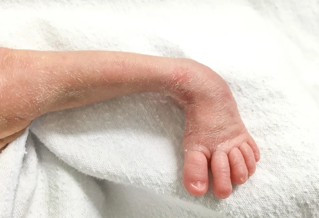


Club Foot In Infants Reasons Signs Remedies


Clubfoot Deformity Talipes Equinovarus
This Pin was discovered by Danielle Brown Discover (and save!) your own Pins on You should ask them the gender again when you get the next ultrasound if the baby is cooperating, it'll only take a sec Hope things go well at the next us!This congenital anomaly is seen in one out of every 1,000 babies, with half of the cases of club foot involving only one foot There is currently no known cause of idiopathic clubfoot, but baby boys are twice as likely to have clubfoot compared to baby girls Neurogenic Clubfoot Neurogenic clubfoot is caused by an underlying neurologic condition For instance, a child born with spina bifida A


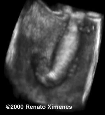
Ssrd Interesting Cases Fetal Clubfoot Ultrasound Image 3d Image


Ssrd Interesting Cases Fetal Clubfoot Ultrasound Image 3d Image
But, she did say that the ultrasound tech noticed baby girl has one club foot I know this can be common &This study suggests karyotyping should be considered even with isolated clubfoot because 111% of isolated cases ended up being complex at birth; Ultrasound Birth Defects Club Foot Club Foot Clubfoot results when the bones and ligaments of the foot do not form like they typically would during a pregnancy, causing them to curl up into a ball, rotate inward, or "club" Clubfoot can be the only defect at birth (isolated clubfoot), or be one of several different features of an underlying genetic syndrome It has many


Normal Fetal


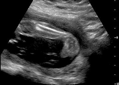
Ultrasound Video Showing Club Foot Fetal Anomaly Scan Youtube
Babies who are born with a foot that's twisted inward and downward have a birth defect called clubfoot Find out what may cause it and how doctors fix it before babiesI am so excited to offer such a unique & special keepsake and transforming your prints & art into a beautiful piece of jewelry or ornaments just for you that will last a lifetime! In babies who have clubfoot, the tendons that connect their leg muscles to their heel are too short These tight tendons cause the foot to twist out of shape Clubfoot is one of the most common congenital birth defects It occurs in about 1 in every 1,000 babies born in the US and affects more boys than girls


What Is Clubfoot And How Is It Treated


Clubfoot Paley Orthopedic Spine Institute
Most of the time, a baby's clubfoot is diagnosed during a prenatal ultrasound before they are born About 10 percent of clubfeet can be diagnosed as early as 13 weeks into pregnancy By 24 weeks, about 80 percent of clubfeet can be diagnosed, and this number steadily increases until birth If a child is not diagnosed before birth, clubfoot can be seen and diagnosed as soon as they are bornEvery 4 to 7 days, your baby's surgeon takes off the cast, moves your baby's foot closer to the correct position and puts on a new cast Before your baby gets his last cast, his surgeon may cut the heel cord (also called the Achilles tendon) This is the tendon that connects the heel to muscles in your baby's calf Cutting the heel cord allows it to grow to a normal length by the time Clubfoot is a birth defect that causes a child's foot to point inward instead of forward The condition is normally identified after birth, but doctors can also tell if an unborn baby has
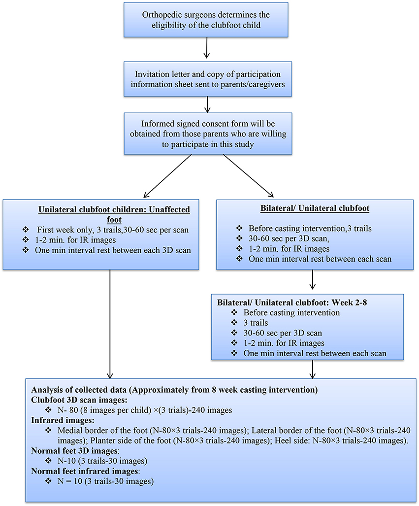


Frontiers Developing A Three Dimensional 3d Assessment Method For Clubfoot A Study Protocol Physiology
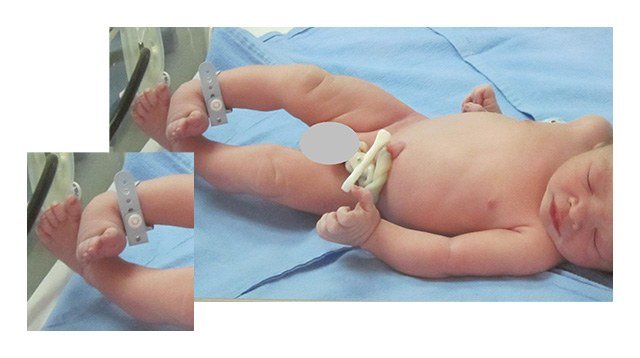


This Dad Is Glad He Got A Second Opinion On His Son S Clubfoot
Club foot tends to be familial in a significant number of cases Wynne Davis supported the polygenic theory and showed a rapid decrease in incidence of clubfoot from first to second to third degree relatives About 29% of siblings in the first degree relatives had this deformity as compared to 12 per thousand masses and chances of getting affected in siblings are more than 25 timesMy 8 month old has 2 club Club Foot Reviewed on Twenty With early treatment, the condition known as clubfoot can be corrected, and your child will be able to walk and run with the best of them What it is A birth defect in which a child's foot points downward and twists inward It can be mild or severe (in severe cases the foot can look as if it's upside down), and it can affect one foot


Clubfoot Johns Hopkins Medicine


Podopeds Clubfoot Flashcards Memorang
Just had our anatomy scan, I'm w5d Doctor said everything looks good & As Dr N explained that the results of the ultrasound showed bilateral club surgery if the baby was in the 5% for whom casting and braces didn't work Dr N inquired if we'd found out the baby's gender, and we said no He said, "I was just curious, as boys are twice as likely to have club foot than girls" We spent the last few minutes of the appointment talking about my health Club foot is usually diagnosed after a baby is born, although it may be spotted during the routine ultrasound scan done between 18 and 21 weeks Melissa trovato, md, william ide, md Clubfoot can have postural and structural characteristics that are classified by the pirani and demeglio scales Club foot can't be treated before birth Club foot may affect one or both feet


What To Expect When Your Child Will Be Born With Clubfeet Living The Diagnosis


Clubfoot Deformity Talipes Equinovarus
Club foot present as an inturn of one foot or both feet Diagnosis The condition can be diagnosed inutero via ultrasound or at birth Visual identification of club foot is all that is needed for diagnosis Treatment There are two treatments currently used to treat club foot – the Ponseti Method and surgery Prenatal ultrasound diagnosis of clubfoot is more accurate in singletons with bilateral clubfoot;The most severe form of clubfoot is characterized by the foot or feet being turned inward and pointed downward When both feet are clubbed, the toes turn toward each other Clubfoot is usually an isolated defect Only about 10 percent of babies with clubfeet have any other associated birth defect There are two general categories of clubfoot – intrinsic and extrinsic Intrinsic
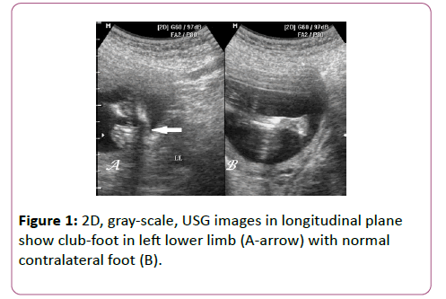


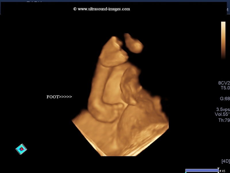
Antenatal 3d Usg In Unilateral Club Foot A Rare Anomaly Insight Medical Publishing


Club Foot Talipes In Babies Causes Signs Treatment Youtube
I have heard some babies look like having club foot on ultrasound and perfectly normal when born I know we wont know anything for sure until further diagnosis but just looking for some positive experiences #1 Bluenpinkmom, Christie11 WellKnown Member Joined Messages 1,162 Likes Received 9 My friend and his wife were told their baby had a club foot Prenatal ultrasound diagnosis of club foot OUTCOME AND RECOMMENDATIONS FOR COUNSELLING AND FOLLOWUP size=1Journal of Bone and Joint Surgery British Volume, Vol 87B, Issue 7, /size Club foot was diagnosed by ultrasonography in 91 feet (52 fetuses) at a mean gestational age of 221 weeks (14 to 356) Outcome was obtained by chart review in


Congenital Foetal Club Foot On Ultrasound In 21week Pregnancy Youtube


Club Foot Nhs


1


Club Foot


Correlations Between Physical And Ultrasound Findings In Congenital Clubfoot At Birth Sciencedirect


Clubfoot Talipes Equinovarus Radiology Key


Clubfoot Deformity Talipes Equinovarus


Clubfoot Wikipedia
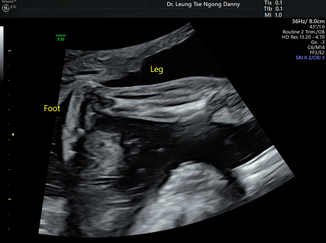


Club Foot Congenital Talipes Equinovarus Hkog Info


Just Had Anatomy Scan What Do Y All Think Clubfoot Forums What To Expect


Clubfoot Versus Positional Foot Deformities On Prenatal Ultrasound Imaging Brasseur Daudruy Journal Of Ultrasound In Medicine Wiley Online Library


Correlations Between Physical And Ultrasound Findings In Congenital Clubfoot At Birth Sciencedirect


Overcoming Clubfoot One Mom S Story Parents
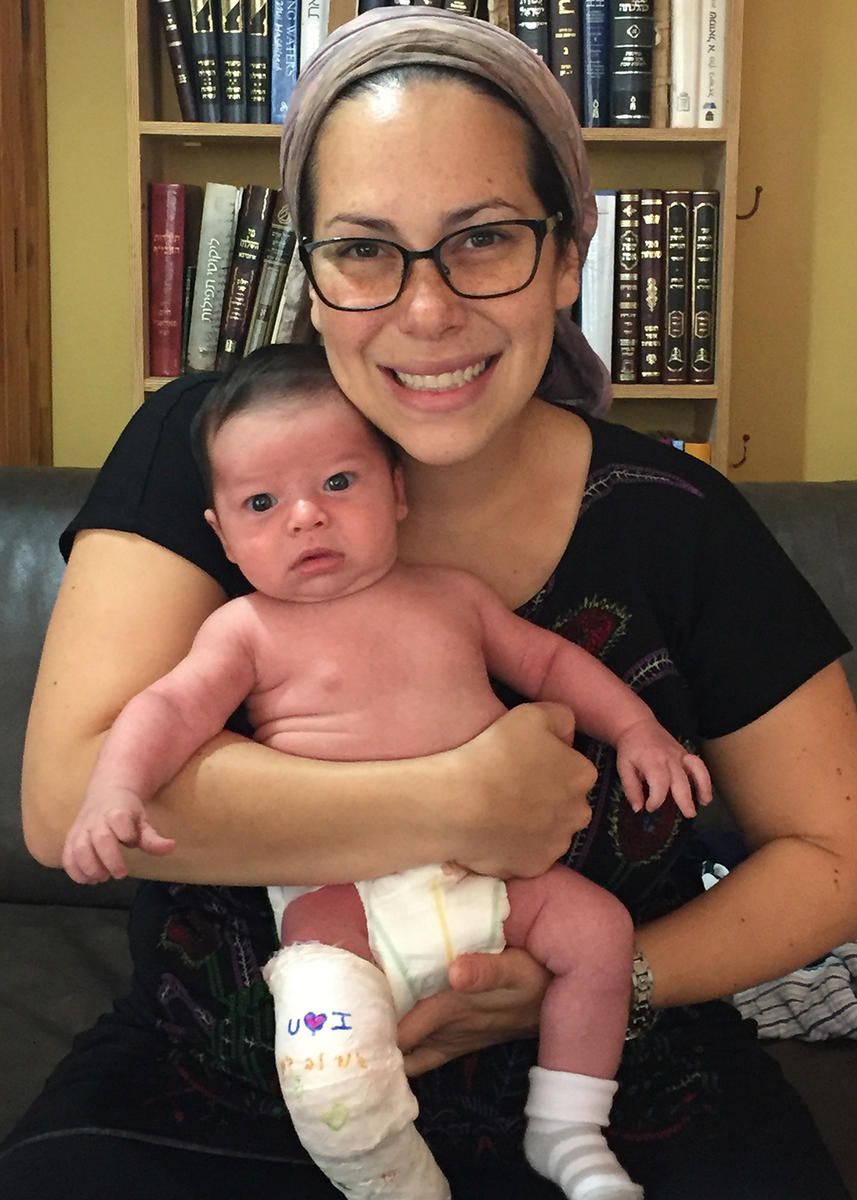


Overcoming Clubfoot One Mom S Story Parents


Value Of The Fetal Plantar Shape In Prenatal Diagnosis Of Talipes Equinovarus Liao 12 Journal Of Ultrasound In Medicine Wiley Online Library


Prenatal Ultrasound Of Case 2 At 34 Gestational Weeks Shows A A Download Scientific Diagram
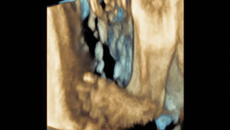


Tackling Talipes Early With A Team Approach Children S Hospital Of Philadelphia


About Clubfoot
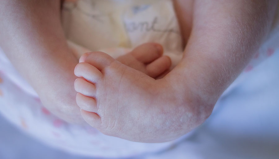


Marlowe S Clubfoot Journey How One Mom Went From Devastated To Reassured Children S Wisconsin


Clubfoot Curved Baby Feet Next Step Foot Ankle Clinic
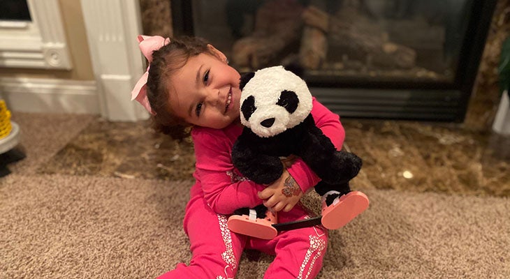


Family Embraces Clubfoot Treatment For Daughter Norton Children S Louisville Ky


Clubfoot Wikipedia


My Journey With Baby S Positional Clubfoot Part 1 Baby Gizmo


Misdiagnosed Clubfoot


Congenital Talipes Equinovarus Radiology Reference Article Radiopaedia Org


The Ultimate Guide To A Clubfoot Baby Simply Working Mama


A Step In The Right Direction Treating Clubfoot Sans Surgery Health Beat Spectrum Health


10 Ut Dms Ideas Medical Ultrasound Ultrasound Technician Diagnostic Medical Sonography


1


404 Not Found Ultrasound Sonography Ultrasound Sonography


The Ultrasound Images Of Fetus Diagnosed As Clubfoot Download Scientific Diagram


Clubfoot Orthopaedia


Clubfoot Boston Children S Hospital


Prenatal Ultrasound Diagnosis Of Club Foot The Bone Joint Journal


Clubfoot Deformity Talipes Equinovarus


Congenital Clubfoot Early Recognition And Conservative Management For Preventing Late Disabilities Springerlink


Clubfeet At 12 Weeks A Transabdominal Scan At 12 Weeks 4 Days Shows Download Scientific Diagram


June 3rd World Clubfoot Day


Club Foot 3d Rendering


Clubfoot Congenital Talipes Equinovarus Pediatrics Orthobullets


Congenital Talipes Equinovarus Radiology Reference Article Radiopaedia Org


A Gallery Of High Resolution Ultrasound Color Doppler 3d Images Fetal Face And Neck


Clubfoot In Children Lurie Children S


Club Foot Antenatal Ultrasound Radiology Case Radiopaedia Org


Clubfoot Causes And Treatments


Three Dimensional 3d Ultrasound Studies A Bilateral Club Foot Download Scientific Diagram
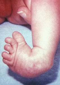


Prenatal Detection Of Clubfoot Key Points The Obg Project


Pdf Congenital Talipes Equinovarus A Case Report Of Bilateral Clubfoot In Three Homozygous Preterm Infants Semantic Scholar


Clubfoot In Newborns Paedicare Paediatricians
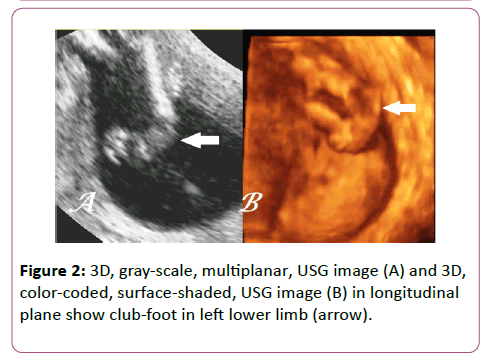


Antenatal 3d Usg In Unilateral Club Foot A Rare Anomaly Insight Medical Publishing


Club Foot


Prenatal Ultrasound Diagnosis Of Club Foot The Bone Joint Journal


Fetus General Normal Fetal Anatomy Ultrasound Services Service Provider From Ernakulam


4d Assessment Of Motoric Function In A Singleton Acephalous Fetus The Role Of The Kanet Test


Ultrasound Video Showing Fetal Anomalies Clubfoot Encephalocele Kyphosis And Placental Mass Youtube


Club Foot


First Trimester Physiological Development Of The Fetal Foot Position Using Three Dimensional Ultrasound In Virtual Reality Bogers 19 Journal Of Obstetrics And Gynaecology Research Wiley Online Library


Clubfoot Healthdirect


Club Foot


Clubfoot Boston Children S Hospital
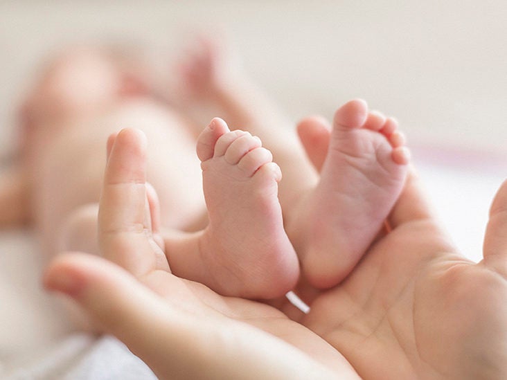


Clubfoot Repair Treatments Procedure Outlook
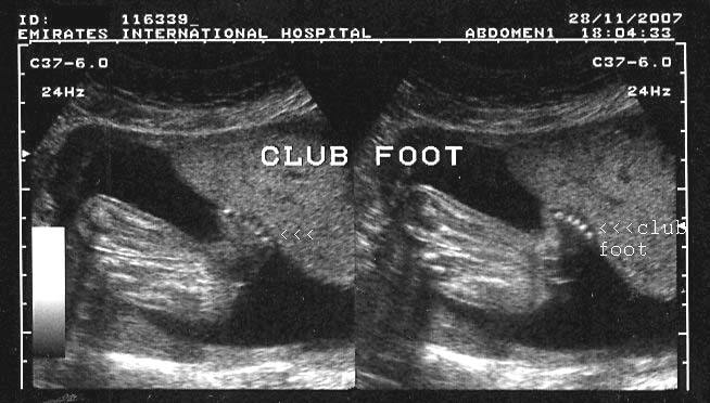


A Gallery Of High Resolution Ultrasound Color Doppler 3d Images Fetal Spine


Clubfoot Symptoms And Causes Mayo Clinic


Clubfoot Versus Positional Foot Deformities On Prenatal Ultrasound Imaging Brasseur Daudruy Journal Of Ultrasound In Medicine Wiley Online Library


A Peachtree City Life An Update On Our Baby Girl Clubfoot


Club Foot


Club Foot In Ultrasound Babycenter


Clubfoot Deformity Talipes Equinovarus


Talipes Equinovarus Club Ultrasound Guided Tips Facebook


Club Foot


Club Foot Talipes Equinovarus Ankle Foot And Orthotic Centre


Kentucky Family Describes Clubfoot Treatment Process Whas11 Com


